
Bone health is often overlooked until something goes wrong. A fracture after a minor fall. Persistent back pain. A gradual loss of height. These are often the first warning signs that bone density may already be declining. The challenge is that bone loss is typically silent. It develops slowly, without symptoms, and many people are unaware there is an issue until significant changes have already occurred.
This is where DEXA bone scanning plays a crucial role. It provides a precise, evidence-based assessment of bone mineral density, helping to identify osteopenia and osteoporosis before fractures happen. But one of the most common questions people ask is simple. Why does a DEXA bone scan focus on the lumbar spine and hip? Why not scan the entire skeleton?
The answer lies in bone biology, fracture risk prediction, and decades of clinical research. These two areas provide the most accurate and clinically meaningful insight into your overall bone health.
What a DEXA Bone Scan Actually Measures
DEXA stands for Dual-energy X-ray Absorptiometry. It is considered the gold standard for measuring bone mineral density. The scan uses very low-dose X-rays to determine how much mineral, primarily calcium, is present in your bones. This measurement reflects bone strength and fracture risk.
Unlike a standard X-ray, which shows structure, a DEXA scan measures density. The result is a precise numerical value that can be tracked over time. This allows clinicians to detect early bone loss, monitor changes, and assess the effectiveness of lifestyle or medical interventions.
The scan produces two key values:
T-score compares your bone density to that of a healthy young adult
Z-score comparing your bone density to others of the same age and sex
These scores help classify bone health into normal, osteopenia, or osteoporosis.
But the accuracy of these scores depends on where the measurements are taken. That is why the lumbar spine and hip are used.
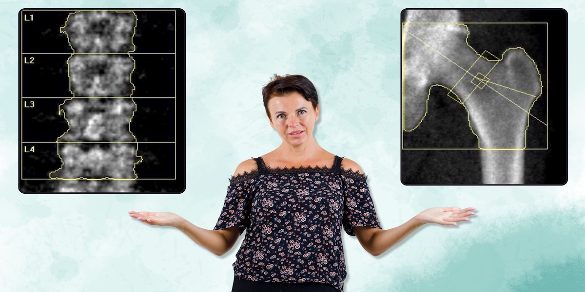
Why the Lumbar Spine Is Scanned
The lumbar spine is located in the lower back and consists of five vertebrae. We scan L1- L4 for a DEXA Bone Scan. These bones are rich in trabecular bone, sometimes referred to as spongy bone. This type of bone is highly metabolically active and remodels more quickly than dense cortical bone.
Because trabecular bone changes rapidly, the lumbar spine is often the first place where bone loss appears. This makes it particularly valuable for detecting early osteopenia or osteoporosis. Hormonal changes, ageing, nutritional deficiencies, and certain medications all affect this region first.
This is especially important for women approaching menopause. Oestrogen plays a vital role in maintaining bone density. When levels decline, bone turnover increases, and trabecular bone can decrease rapidly. The lumbar spine measurement captures this early shift.
The lumbar spine is also extremely useful for monitoring treatment. Because it responds quickly to change, improvements in bone density can often be seen sooner than in other areas. This makes it ideal for tracking progress following lifestyle changes, supplementation, or medication.
In simple terms, the lumbar spine acts as an early warning system. It helps identify bone loss before it becomes severe.
Why the Hip Is Scanned
While the lumbar spine detects early bone loss, the hip plays a different role. It is one of the most clinically important fracture sites. Hip fractures are associated with reduced mobility, loss of independence, and increased health complications.
Because of this, hip bone density is one of the strongest predictors of future fracture risk. Measuring this area helps identify individuals who may be at risk of serious injury.
The hip contains a combination of trabecular and cortical bone, making it a balanced site for assessment. It is also less affected by degenerative changes that can influence spine readings, particularly in older adults. Conditions such as osteoarthritis or calcification can sometimes artificially elevate lumbar spine results. The hip provides a stable comparison.
Another advantage is consistency. Hip measurements are highly reproducible, making them ideal for tracking bone density over time. This allows clinicians to monitor trends and detect gradual changes.
The hip measurement is also used in fracture risk calculations. These tools estimate the likelihood of fractures over time and help guide clinical decisions.
Why Both Areas Are Scanned Together
Scanning both the lumbar spine and hip provides a comprehensive assessment. Each site offers different information.
The lumbar spine detects early bone loss.
The hip predicts fracture risk.
Together, they provide the most complete picture of skeletal health.
This dual-site approach is recommended by international osteoporosis guidelines. It improves diagnostic accuracy and reduces the chance of missing bone loss.
Sometimes results differ between the two sites. For example, someone may have reduced spine density but normal hip density. This may indicate early bone loss. Alternatively, the hip may show lower density, highlighting fracture risk.
Using both measurements ensures that bone health is assessed thoroughly.
How These Areas Reflect Whole Body Bone Health
Bone loss is typically systemic. This means when bone density declines, it tends to decline throughout the skeleton. The lumbar spine and hip act as representative sites because they contain different bone types and respond to different physiological changes.
Research has shown strong correlations between bone density at these sites and fracture risk in other areas such as the wrist, shoulder, and pelvis. This is why scanning the entire skeleton is not necessary.
By focusing on the most informative locations, DEXA scanning provides accurate results with minimal radiation exposure.
Who Should Consider a DEXA Bone Scan
DEXA bone scans are not just for older adults. Many factors influence bone density, including lifestyle, nutrition, hormones, and genetics.
You may benefit from a DEXA bone scan if you:
- Are approaching or experiencing menopause
- Have a family history of osteoporosis
- Have low body weight
- Follow restrictive diets
- Are an endurance athlete
- Have experienced stress fractures
- Take long-term steroid medication
- Have low vitamin D levels
- Have thyroid or hormonal imbalances
- Want to monitor bone health proactively
Early testing allows for early intervention.
Lifestyle Factors That Affect Bone Density
Bone is living tissue that responds to lifestyle choices. Positive factors include:
- Resistance training
- Weight-bearing exercise
- Adequate protein intake
- Sufficient calcium intake
- Vitamin D exposure
- Balanced calorie intake
Negative influences include:
- Low-calorie diets
- Excessive endurance training
- Smoking
- Alcohol excess
- Hormonal imbalances
- Rapid weight loss
These factors often affect the lumbar spine first, reinforcing why it is scanned.
Why Early Detection Matters
Osteoporosis is often called a silent condition. There are usually no symptoms until a fracture occurs. By that point, bone density may already be significantly reduced.
DEXA scanning helps identify bone loss early. This allows individuals to take action before fractures happen. Many people can improve bone density with lifestyle changes alone.
Monitoring also provides reassurance. Tracking bone density over time helps determine whether interventions are working.
How DEXA Results Are Used
After your scan, your report will typically include:
- Lumbar spine bone density
- Hip bone density
- T-score
- Z-score
- Classification of bone health
- Fracture risk indication
These results help guide next steps. This may include:
- Nutrition advice
- Strength training recommendations
- Supplement guidance
- Hormone evaluation
- Follow-up scanning
The goal is prevention, not just diagnosis.
Frequently Asked Questions
Why not scan the wrist instead?
The wrist is sometimes used when spine or hip scanning is not possible. However, it is less predictive of fracture risk and not considered the gold standard.
Why not scan the whole body?
Whole-body scans provide additional information but are not necessary for bone density diagnosis. The spine and hip provide the most clinically relevant data.
How often should I have a DEXA scan?
This depends on your results. Typically, every one to two years if monitoring is needed. Some people may scan less frequently.
Is the scan safe?
Yes. Radiation exposure is extremely low, much lower than a standard X-ray.
Can bone density improve?
Yes. With the right nutrition, resistance training, and medical support, bone density can stabilise or improve.
The Gold Standard for Bone Health Assessment
The reason DEXA scans focus on the lumbar spine and hip is simple. These areas provide the most accurate and clinically meaningful assessment of bone health.
The lumbar spine highlights early bone loss.
The hip predicts fracture risk.
Together, they reflect the strength of the entire skeleton.
This approach is backed by research, recommended by guidelines, and used worldwide.
Take Control of Your Bone Health
Bone health affects strength, mobility, and long-term independence. A DEXA bone scan provides clarity. By scanning the lumbar spine and hip, you gain a precise understanding of your bone density and fracture risk.
Early detection allows you to take action. Whether that means improving nutrition, increasing strength training, or monitoring changes over time, the information empowers you to make informed decisions.
Your bones support you every day. Understanding their health is one of the most valuable investments you can make for your future.